by Jonathan N. Roth, Ph.D., Micrology Laboratories L.L.C.
Certain compounds are not uncommonly incorporated into media formulations due to their unique property of changing color when they change from the reduced state to the oxidized state, or vice versa. These compounds (herein referred to as Redox Compounds) may include various chemical makeups, and examples include such compounds as methylene blue, Resazurin, and tetrazolium salts.
Methylene Blue is commonly incorporated in anaerobic media in order to signal when the medium is no longer in its anaerobic (oxygenless) state. Such media are generally without dissolved oxygen when first removed from a sterilized (autoclave) and so are colorless (the methylene blue is reduced). But as they cool and are exposed to the normal atmosphere, they will absorb oxygen and turn a blue color(the methylene blue becomes oxidized). Therefore, the appearance of blue color signals the loss of the anaerobic condition of the medium.
Resazurin goes from the blue oxidized state to stages of purple to pink and finally colorless as it is reduced. Both it and methylene blue have been used in the determination of milk quality by determining how long it takes for milk containing one of the dyes to go from a blue to a white condition. In this test method, the shorter the reduction time, the higher the population of active microbes is considered to be in the milk.
Another group of Redox compounds used in microbiological media consists of various Tetrazolium Salts, with the commonest being 2,3,5-Triphenyl-2H-Tetrazolium Chloride (TCC). TCC is colorless when in the oxidized state, but assumes a bright red color when reduced. TCC is used as a necessary ingredient in some microbiological media such as Hach’s ColiBlue® media, some of 3M’s Petrifilm® formulations, where the visibility of some microbes would be lacking without the red color imparted to them by the TCC. Micrology Laboratories utilizes TCC in some of its formulations both to impart enhanced contrast and visability of microbial growth to inexperienced students, but more importantly to differentiate and separate microbial colony growth from other materials (debris) that may be present in a test sample. Most bacteria growing in an aerobic atmosphere will cause the reduction of TCC with attendant red bacterial colony color. Therefore, its use is only to enhance the visability of the bacterial growth and not to differentiate various species of bacteria.
It should be noted that care must be exercised to avoid too high TCC levels in media as this can result in the inhibition of some bacterial species. If the TCC is used judiciously, there should not be a statistically significant reduction in the bacterial counts between TCC containing media and corresponding formulations without TCC .
Specific example of TCC use in a medium to differentiate and enhance the contrast of bacterial colonies from surrounding debris: superior visibility and quantification of bacterial presence in meat products
A major problem in the determination (quantification) of bacterial populations in/on meat products is the fact that in order to achieve satisfactory counts, a relatively large sample of meat must be blended (thoroughly macerated) to make a homogenous solution where-in any bacteria or Colony Forming Units (CFUs) are separated from the meat substrate and available to grow into individual colonies when the sample is plated out and incubated. For example, 10 grams of meat may be blended in 90 mL of sterile diluent. A given amount of this blended solution (commonly 1-5 mL) is mixed with a nutrient microbiological medium and plated out in a petri dish (“pour plate”). The real problem is that the sample which is mixed with the medium contains large numbers of small meat fragments which are visible either to the unaided eye or under 10X magnification. Also, it is virtually impossible to extract all bacterial cells or aggregates from all meat fragments.
When the “pour plate” is incubated and bacterial colony growth occurs, it can be very difficult to differentiate bacterial colonies from meat fragments. They both appear as white objects on a dark background when illuminated from the top. The experienced technician can generally distinguish the round, uniform bacterial colony from the irregular, non-uniform meat fragment, but it is an eye straining, tiresome and time consuming job. Also, often it is virtually impossible to determine if there is any bacterial growth present in or on the meat fragments themselves. These problems can result in serious miscalculations of the actual numbers of bacteria present in the test sample. In the case of overly zealous technicians, the counts result in very high false positives, and in the opposite case there may be very high false negatives, neither of which is desirable. Both scenarios can be avoided by using Easygel with TCC as produced and available from Micrology Laboratories.
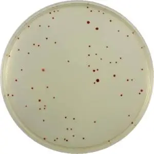
The beauty of the Easygel/TCC method is that the TCC in the medium will be colorless until bacterial colonies are formed and effect reduction of the TCC, coloring the colonies with a bright red center. The meat fragments in the medium will not cause any TCC reduction and so they remain as white, uncolored pieces of debris. However, additionally, if there are any bacteria embedded on or in a meat fragment, they will grow and a red spot (colony) will appear on the fragment (it is very unlikely that such growth would be detected by even the most experienced technician without the TCC help).
Total Count (Standard Plate Count) and Tryptone Soy Easygel formulations from Micrology Laboratories are available and suited for use with Pretreated Petri Dishes containing TCC in their treatment layer on the dish bottom. Either is an excellent choice for total heterotrophic plate counts. Use of the Easygel Method is simple, saves time, and is inexpensive.